
BPS Parent Guide
Medically reviewed and edited by Marisa Schwab, MD
Written by Emily Lake, PhD
Last updated 09/18/2025
What is BPS?
The cause of BPS is unknown and nothing you did or didn’t do in pregnancy caused it.
BPS is a benign (non-cancerous) mass of lung tissue, normally affecting just one lung lobe. BPS is a type of congenital lung lesion (CLM) or malformation. It is not a cancer or a tumor. The mass develops in the lungs very early during pregnancy, sometime before the 10th week of gestation. The BPS mass is almost always solid. The lung tissue that is affected doesn’t work normally, but the rest of the lung—and the other lung lobes—usually develop and function well.
The main thing that distinguishes a BPS lesion from CPAM is that a BPS gets its blood supply from another systemic vessel outside the pulmonary (lung) system. This is commonly, but not always, the aorta.
Like CPAM, most BPS lesions will shrink before birth and the majority of babies are asymptomatic, meaning they have no breathing problems before and after delivery. If surgeons recommend that the BPS and affected lung lobe (in the case of intralobar sequestrations) be removed after birth, the healthy lung tissue will expand to fill the space. The vast majority of kids will have no long-term impacts on their health or activity levels.
Are there different types of BPS?
The BPS can be intralobar, meaning the mass is part of a lung lobe (but with an external blood supply), or extralobar, meaning the mass is outside of the lung lobes. Intralobar sequestrations are thought to be more common than extralobar sequestrations. In about a quarter of cases, the lung mass will show characteristics of both CPAM and BPS (typically a cystic mass with an external blood supply). These are called hybrid lesions.
Imaging before birth gives doctors an idea of what the lesion looks like, but it won’t be until a CT scan is done after birth that they’ll have a good idea of whether this is a BPS or a different type of congenital lung lesion. It will also give a better idea of the location and characteristics of the BPS.
How rare is it?
Congenital lung malformations, including BPS, are uncommon. Estimates for all lesions are 1 in 2,500 live births. BPS is the second most common congenital lung malformation after CPAM. Currently, approximately 70% of babies with a congenital lung malformation are diagnosed before birth.
Is there a risk of cancer?
There is not thought to be any risk of cancer from a BPS lesion. There may be an increased risk of cancer with CPAM, and some patients have hybrid lesions, meaning both sequestration and CPAM.

A sketch of the lungs with a BPS lesion (shaded) showing its blood supply outside the lungs.
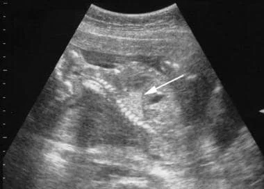
An intralobar BPS lesion viewed on an ultrasound. The lesion appears brighter than the surrounding healthy lung tissue.
Source: Medscape article
The BPS can be intralobar, meaning the mass is part of a lung lobe (but with an external blood supply), or extralobar, meaning the mass is outside of the lung lobes
What happens during a BPS pregnancy?
Your pregnancy will be more closely monitored than an uncomplicated or lower risk pregnancy. Your medical team will look at the size and appearance of the BPS and track its growth over time. You will be asked to come in for ultrasounds more frequently. Depending on the size, this could be weekly or every couple of weeks. In rarer high-risk cases, you might have to come in twice a week. Sometimes a fetal MRI may be carried out during the pregnancy to get a more detailed picture of the BPS, although this is not offered everywhere or for every case. In certain cases, especially where the heart is pushed or compressed by the BPS, a fetal echocardiogram may be obtained to get a more detailed look at blood flow and how the heart is functioning. Ultrasounds can normally be carried out where you were already receiving prenatal care. You will need to go to a specialist hospital for the fetal MRI and fetal echocardiogram.
Who will be involved in my care?
Many different specialists will now be involved in you and your baby’s care. Depending on where you’re receiving prenatal care, you will be connected to a maternal-fetal medicine (MFM) specialist. This is a doctor who works with pregnant women and unborn babies during high risk and complicated pregnancies. The MFM should understand congenital lung malformations and will make sure you and your baby are properly cared for. A radiologist will take specialized images of your unborn baby during ultrasounds and interpret these findings. They will be looking for how large the BPS is and whether it shows any characteristics that might change the course of management and treatment. They will also closely look for any signs that the BPS might be pushing on other organs, such as shifting the heart to the other side of your baby’s chest (mediastinal shift). Doctors will also look for hydrops, which is when fluid builds up in a body’s tissues or organs (such as the lungs, heart, or belly). Closer to the third trimester you will then have a consultation with a pediatric surgeon to understand if they plan to operate, when this would be, and what an operation would look like. You may also ask for a consultation with a neonatologist (the specialist doctors who work in NICUs) to understand what will happen at birth if your baby is symptomatic.
What are the risks during pregnancy?
The risks depend on the size, type, and other findings on imaging (such as mediastinal shift and hydrops). Most lung malformations increase in size between 20 and 26 weeks gestation, and then will stay the same or decrease in size.
The CVR Number
The most common way to measure the size of the lesion is the CPAM Volume Ratio (CVR), which estimates the BPS size in relation to the baby’s head. This gives doctors an idea of how large the BPS is and, therefore, whether there is a risk of hydrops. In general, a CVR of less than 1.6 is considered lower-risk, while a CVR of greater than 1.6 should lead to increased monitoring because of the higher chance of developing hydrops. The vast majority of CPAM babies without hydrops do very well.
In some countries, doctors will tell you the CVR number at each appointment. In others, they won’t share this information. Some parents find it useful to know and track the CVR while others find that it adds too much stress. Remember that the CVR is an estimate and not 100% accurate. The CVR will change depending on the baby’s position that day, where the cursor took the measurement from, and who the ultrasound tech was. Because these are often very small numbers, a millimeter one way or the other can make a big difference to the CVR. Some parents ask if they can see the same ultrasound technician for every appointment to control for variation in how people take measurements, but this isn’t always possible.
If your doctor has not shared the CVR number with you, you can calculate it using our calculator if you have the measurements of your baby’s BPS (length, height, width) and head circumference.
High risk cases
Higher risk cases are uncommon. If a baby’s BPS is very large (greater than a CVR of 1.6) they will be watched very closely for signs of hydrops or other fetal distress. Hydrops can be dangerous for both mother and baby, so your doctors will watch you very closely. Depending on the size and change of the CPAM, your doctors may recommend a treatment during pregnancy.
Maternal steroids (betamethasone) may help shrink the BPS and can prevent or reverse hydrops in some cases. You may be given multiple doses of steroids.
Fetal surgery and EXIT procedures are rarely considered and carry significant risks.
The CVR will change depending on the baby’s position that day, where the cursor took the measurement from, and who the ultrasound tech was.
What to expect after birth
Your doctors will talk to you about what they expect for your baby, based on your prenatal imaging. Most babies with BPS are asymptomatic: they breathe normally at birth and need only routine care. If your prenatal imaging has been reassuring, your team may let you deliver at the hospital of your choice and evaluate your baby in the clinic. Other patients with higher risk need to deliver at a hospital with a NICU, in case your baby needs medical care after delivery. How your baby looks after delivery will determine if the baby goes to the NICU or can be with you in your room.
All babies get a chest X-ray a few hours after birth, even if they’re breathing normally. The chest X-ray often looks normal without a visible lung malformation, even though it’s still there. If your baby doesn’t have any symptoms, the next step after you go home will be a CT scan of the chest (usually when they’re a few months old) and a visit with the pediatric surgeon. The CT scan is important because it shows the location and detailed characteristics of your baby’s BPS. The surgeon will talk to you about whether they recommend surgery, and what that looks like.
If your baby has symptoms, which happens in about 10% of cases, they may need surgery shortly after birth. The surgeons and neonatologists will talk to you about the details of surgery.
Management and treatment
There are two main paths for BPS management and treatment. Which one you choose depends on the specifics of your baby’s BPS, the advice of your medical team, where in the world you are located, and what you feel is best for your child.
Elective surgical removal (most often a thoracoscopic or open lobectomy) in later infancy— normally around 3–6 months—even if a child has no symptoms. This means that the surgeon will remove the entire affected lung lobe or, in extralobar cases, the surgeon will normally just remove the sequestration without removing any lung lobes. This is the common treatment path in the USA and usually recommended for all cases of intralobar sequestrations, even if there are no symptoms. The reason this is recommended by most doctors in the USA is to prevent repeated and severe infections (such as pneumonia) for the rest of the patient’s life and make the diagnosis certain.
The recovery time depends on whether the operation is done via the open or minimally invasive technique. After a thoracoscopic lobectomy, babies usually recover quickly and spend one or two days in the hospital. If a lobe has been removed, the remaining lung will expand and grow to compensate for the removed lobe, and most children will have the same lung function as someone who didn’t have lung surgery as a baby. They’ll be able to do all the activities and sports they want when they grow up.Careful observation (“watchful waiting”) with scheduled check-ins and regular imaging. This is the common treatment path for patients with asymptomatic BPS (especially extralobar sequestrations) in Canada, Australia, and many European countries. If symptoms develop—recurrent chest infections, wheezing not explained by common causes, or a spontaneous pneumothorax—surgery is then usually recommended.